Dental X-rays are one of those things a lot of patients worry about before they ever step into a dental chair. You hear the word “radiation” and your mind jumps to big cancer scans or airport security machines. But in modern dentistry, dental x-ray safety isn’t something to be afraid of — it’s something to understand. These images, known as dental radiographs, give your dentist information that can’t be seen with the naked eye, and that often makes the difference between catching a problem early and treating a big, painful surprise later.
What Dental X-Rays Actually Are
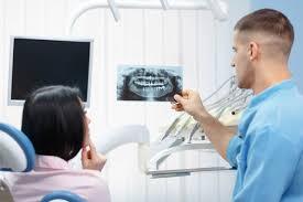
Dental X-rays are quick photographic images made using controlled bursts of radiation to penetrate bone and teeth. The structures show up in contrast: enamel and dense bone appear lighter on the film, while decay, infections, and soft tissues show up darker. Dentists use several types — bitewings to check for small cavities between teeth, periapical radiographs to see up and down a single tooth, and panoramic X-rays that scan the entire dental arch at once. Each type has its purpose based on what your mouth needs.
They’re a crucial diagnostic tool during routine exams, but they’re not taken at every single visit unless there’s a reason. Modern radiographs are digital, which means exposure is very low and images can be seen instantly on a computer screen.
Why You Might Need X-Rays During an Appointment
Dentists don’t order X-rays just to follow a habit. The decision about when need X-rays depends on your oral health history, your age, and what the dentist suspects during an exam. If you’re a new patient with no recent images, the dentist might start with a full set to establish a baseline. If you have a lot of fillings, crowns, gum disease, or a history of decay, you might need them more often — maybe every six to twelve months — because radiographs help monitor old problems and catch new ones.
Children and teenagers often need images more frequently than adults, because their teeth and jaws are developing rapidly. Dentists use X-rays to check how permanent teeth are coming in, whether baby teeth need extraction to make space, and if there are hidden decay spots. Adults with certain risk factors might also require more frequent checks. Radiographs are also important before major procedures — implants, braces, root canals, or difficult extractions — because they let the clinician plan safely and avoid surprises.
How Safe Are Dental Radiographs Really?
Safety is the question most people ask first. Dental X-rays use ionising radiation, but in amounts so low that they’re considered safe for most people. The exposure from a standard bitewing radiograph is a tiny fraction of what your body absorbs from natural environmental radiation every day. Even when compared with other types of medical imaging, dental radiographs represent a very small dose.
The American Dental Association and other health authorities recommend that X-rays be taken only when clinically necessary, and that radiation exposure be kept “As Low As Reasonably Achievable,” a principle known as ALARA. Protective equipment like lead aprons and thyroid collars, combined with digital imaging that uses far less radiation than old film technology, keeps your dose even lower.
Even though radiation is involved, for most adults and children the risk of harm from dental X-rays is considered extremely low. Dentists are trained to evaluate both benefits and risks before recommending an image. If you ever have concerns — for example, if you’re pregnant — sharing that with your dentist helps them take extra precautions or choose alternative methods when appropriate.
Benefits of Using Dental Radiographs in Care
Without radiographs, a dentist might miss decay hidden between teeth, infections in the bone, or early signs of gum disease. Many cavities start in places you can’t see with a mirror, and they don’t show symptoms until they’re already advanced. Radiographs let dentists diagnose these problems early, so treatment can be simpler, cheaper, and less painful.
Radiographs also help evaluate bone health around the teeth, which is especially important for people with periodontal issues. Small bone losses that aren’t obvious during a visual exam can show up clearly on an X-ray. For orthodontics, they show how teeth are aligned deep within the jaw. For wisdom teeth, they show position and development. For implants, they assess bone density and space. Without these images, treatment planning is guesswork at best.
Frequency and Individual Needs
There’s no one-size-fits-all answer for when need X-rays. Healthy adults with strong oral hygiene and no history of decay may only need images every couple of years. People with a history of dental issues, gum recession, or high sugar diets could need them more often, sometimes every six to twelve months. Your dentist customises the frequency based on your risk factors and clinical findings.
Children and teens usually need them more often because their mouths change so quickly. For example, before orthodontic planning, panoramic radiographs show a full view of developing teeth and jaw structures. This helps guide decisions about braces or extractions.
Addressing Common Misconceptions About Safety
There’s a persistent myth that dental X-rays cause cancer or significant harm due to radiation. In reality, the small amount of radiation used in dental settings is so minimal that it’s not considered harmful by major health organisations. Modern digital technology further shrinks exposure, and protective gear limits radiation to areas that don’t need imaging.
Another misunderstanding is that X-rays are taken routinely at every check-up. They’re not. Most dentists follow evidence-based guidelines, meaning radiographs are only recommended when there’s a clinical need — such as suspected decay, ongoing treatment, or monitoring of a known issue. You can always ask your dentist why a particular type of image is being recommended, and how it contributes to your care.
Patient Awareness and Communication
If you’re uneasy about radiation or simply curious about dental radiographs, talk to your dentist about what they expect to learn from the images. Good communication helps you understand why they’re recommending X-rays, how often you’ll need them, and what the benefits are for your particular situation. Informed patients tend to feel more comfortable with the process, and they’re more likely to stay on top of preventive care that keeps tiny problems from ballooning into major ones.
Understanding dental X-ray safety and necessity removes a lot of the fear around them. When you see that the exposures are low, that they’re only taken when needed, and that protective steps are in place, it’s easier to view these radiographs as a valuable part of keeping your smile healthy and preventing severe dental problems down the line.